Electroencephalography(EEG) Electrode Placement Navigation System
Electrode Navigation System with high repeatability.
Neurorehabilitation and EEGs are conducted repeatedly over a long period of time for evaluating recovery. In this regard, the repeatability of the EEG electrode placement is one of the most important prerequisites for obtaining reliable EEG signals
Conventional methods for positioning electroencephalography (EEG) electrodes according to the international 10/20 system are based on the manual identification of the principal 10/20 landmarks via visual inspection and palpation, inducing intersession variations in their determined locations due to structural ambiguity or poor visibility.
Therefore, we propose an electrode navigation system to assist in reliable EEG electrode placement, deploying the concept of surgical navigation, which provides real-time image guidance on the position of surgical instruments relative to the patient’s anatomy during surgery. The location of the EEG electrodes is displayed in real time relative to their goal positions, i.e. 10/20 landmark locations in the three-dimensional (3D) virtual space, facilitating electrode placement with ease and precision.
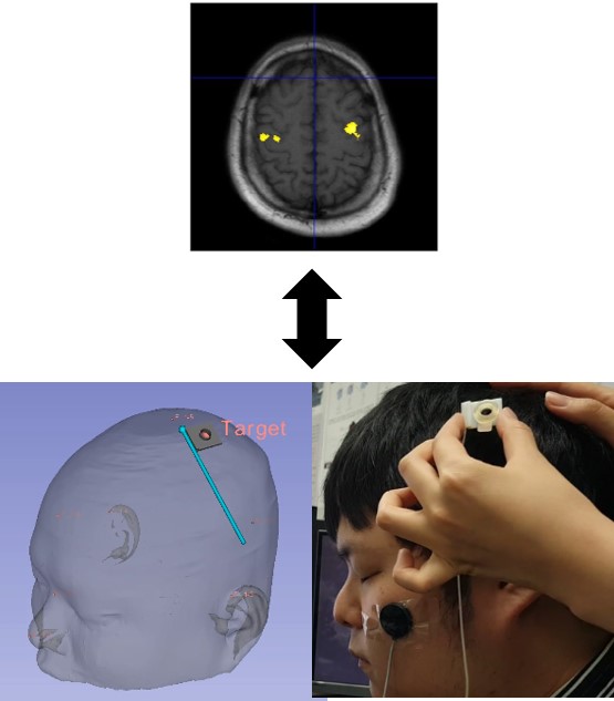
Region of Interest(ROI) based Electrode Navigation System
Conventional EEG electrode placement system does not guarantee of reliable signal for brain-damaged patients that translate the activity area by brain plasticity.
Therefore, the proposed system suggests to placing electrode on ROI determine from fMRI/MRI where brains activity really on. The proposed system provides the visual information on the current location of the EEG electrode with respect to the patient head and ROI from fMRI to track the patient and the EEG electrode in real time with optical tracking system. The patient with ROI and EEG electrode are visualized as 3D models in the virtual space.

Related papers in our group
[1]
Jeon S, Chien J, Song C, Hong J, A Preliminary Study on Precision Image Guidance for Electrode Placement in an EEG Study,Brain Topogr, DOI: 10.1007/s10548-017-0610-y, Dec 2017.