Surgical Navigation System for Vascular Intervention
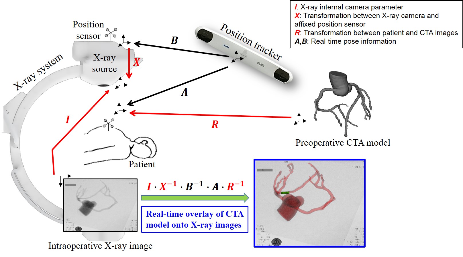
A surgical navigation system is presented to improve the safety and precision of conventional vascular interventions. The proposed system visualizes in real time vascular anatomy or occlusion that are originally invisible in X-ray images and the location of surgical instruments is displayed in the 3D CT model.

Researchers
Sangseo Jeon
System configuration
Component #1
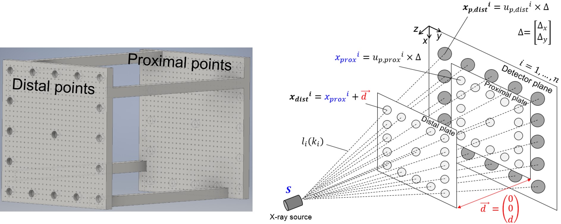
● Geometric X-ray calibration
Geometric X-ray calibration determines internal imaging parameters of cone-beam X-ray scanner and a transformation between coordinate systems of an external position sensor and X-ray source. The derived information is used to reproduce virtual X-ray imaging geometry with respect to the current location of the patient.
Component #2
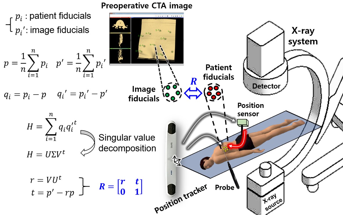
● Patient-to-image registration
Patient-to-image registration is a process to determine a transformation between a preoperative CT image space and the real world represented by a patient-affixed position sensor using artificial fiducial markers or anatomical landmarks. Via this process, real-time movement of the patient can be compensated in the navigation display.
Component #3
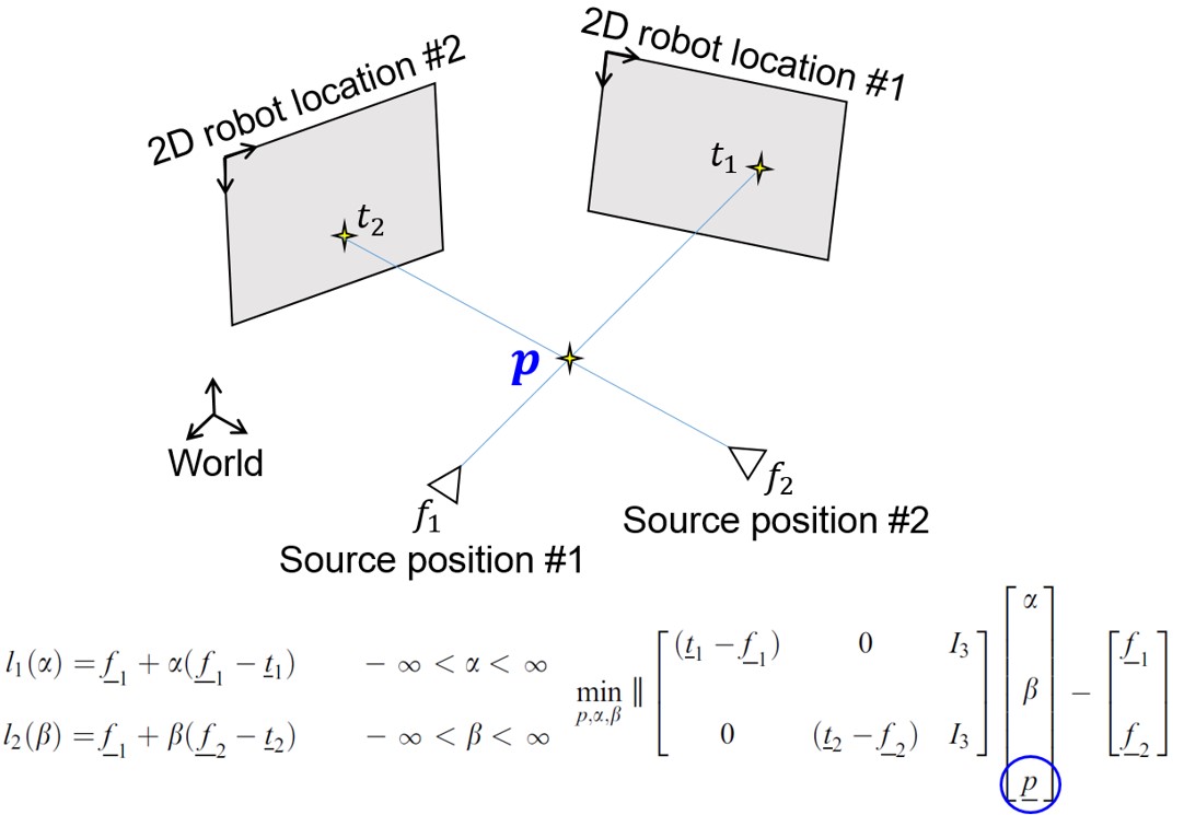
● Instrument localization
With more than two X-ray images and the corresponding X-ray source locations reported from the position sensor, 3D location of surgical instruments can be reconstructed. Following patient-to-imager registration, the 3D instrument location is displayed with respect to the CT image space.